
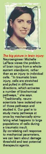
Think
of it as a modern-day version of the canary in a coal mine—
living tissue interfaced with microchip circuits that can function as
an early-warning system for chemical or biological warfare. Biomedical
engineer Steve DeWeerth has been working on the biosensor concept for
years. Now he’s turning it up a notch.
This kind
of research is why biomedical engineering exists,” says DeWeerth,
an expert in computation and neural systems in the Wallace H. Coulter
Department of Biomedical Engineering at Georgia Tech/Emory. “Blending
medicine and engineering allows us to do things that would be impossible
through either specialty alone.”
DeWeerth
is developing two types of biosensing devices, one connecting muscle cells
to electronic circuits to detect effects of chemical or biological agents,
and the other using “cycling probe” technology to match the
DNA fingerprint in air particles or fluids to known viruses or bacteria.
“The
first line of defense against biological and chemical warfare is your
body,” says DeWeerth. “When you get sick, you know there’s
a problem. But we don’t want people to get sick, we want devices
to get sick. It’s like a tiny smoke alarm.” DeWeerth expects
that either device could be ready for use as a building sensor in the
near future.
Devices like
these illustrate the clear relevance of “translational” research,
a new buzzword for research easily translated into benefit for people—and
a concept that has been the compelling force behind biomedical engineering
since its very beginning. You can hear the excitement it generates in
DeWeerth’s immodest but animated view of his department’s mission:
“In all honesty,” he says earnestly as he woos a faculty recruit,
“we’re trying to change the world!”
Immodesty
aside, in its five short years of existence, the Coulter Department of
Biomedical Engineering (BME) has indeed been on a breathtakingly rapid
rise toward the top of its field––accumulating major funding,
top-notch (and enthusiastic) faculty, brilliant students, and sparkling
new facilities. BME also has earned sixth-place ranking from US News &
World Report, surpassing a number of well-respected and established programs.
Despite these
early successes, BME faculty say they plan to stop at nothing short of
creating the gold standard of research and education in biomedical engineering.
The key to creating groundbreaking biomedical technology within the next
100 years, they believe, is to obliterate forever the rigid distinction
between engineering and medicine and establish an entirely new culture
of scientists. This new breed of scientists will be comfortable and conversant
in both medicine and engineering, applying engineering principles to medicine
and using living molecules to design replacements for damaged organs or
tissues.
“Thirty years ago, if someone had said ‘bioengineering’, you might have thought ‘biotechnician’ and pictured a guy who fixes your TV set in the hospital,” quips DeWeerth, “just as 100 years ago, people would have asked, ‘What’s electrical engineering?’ Just look at the way doctors were trained at the beginning of the past century. Fifty years from now, we will reflect on the way physicians and engineers were trained separately and see the distinction as quaint. Biomedical problems need a common solution that uses the knowledge of both medicine and engineering.” (BACK TO TOP)
Pivotal
relationships
One person
who has already seen a great deal of blurring of the distinction between
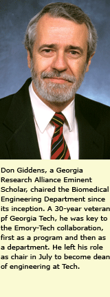
medicine and engineering in a relatively short time is former BME chair
Don Giddens. A 30-year veteran of Tech and a former dean of engineering
at Johns Hopkins, Giddens has been a fulcrum in the relationship between
Emory and Tech for more than 15 years. Since that time, he has seen a
program transpose into a department, propelled by major funding from the
Coulter and Whitaker foundations (see timeline, page 22). Those gifts
are providing precious space to make room for an even more priceless commodity
in the form of faculty and students.
Giddens says another architectural linchpin of the department is the Georgia Research Alliance (GRA), a partnership of Georgia’s research universities, business community, and state government created in 1990. Giddens, who is a GRA Eminent Scholar, says the Emory-Tech collaborations fit perfectly into the GRA’s model of university partnership, as does their heavy emphasis on translational research and research commercialization. He adds that the GRA recently attracted another eminent scholar to BME, Xiaoping Hu, a pioneer in functional magnetic resonance imaging (MRI), who will head a new biomedical imaging technology center at Emory. (BACK TO TOP)
Imagine
these images
The new GRA
scholar will join an already strong and growing medical imaging group
that includes Allen Tannenbaum, an engineer/mathematician whose inventions
are designed to make diagnostic methods less invasive and more effective.
Neurosurgeons
at a few teaching hospitals are beginning to use MRI software developed
by Tannenbaum and his colleagues that visually slices the brain into more
than 120 pieces using “interventional magnetics.” Within seconds,
the slices are reconstructed into a 3-D image that illustrates the exact
location and parameters of a brain tumor. Other software developed by
Tannenbaum virtually flattens the brain onto a sphere, allowing visual
access to the brain’s deep indentations.
Tannenbaum’s
new system of virtual colonoscopy could be a major breakthrough in circumventing
real colonoscopy—“one of the most popular procedures on the
face of the planet,” he says. He believes this system is an improvement
over another virtual colonoscopy system currently being tested at centers
nationwide, including Emory, in which the radiologist visually scans a
series of CT images, searching for the presence and location of polyps.
The drawback, says Tannenbaum, is that radiologists must still do the
searching themselves.
“Imagine
if you could image the colon, reconstruct it virtually, then use that
image to locate polyps,” says Tannenbaum. Imagine no more. Tannenbaum’s
software cuts the colon open virtually, rolls it out like a ceremonial
red carpet, then flattens it into a colorful, cartoon-like image before
the eyes of a radiologist. The computer scans the virtual colon and highlights
suspected polyps in green, allowing the radiologist to correlate the image
with the original CT data and confirm the need—or not—for a
real colonoscopy.
Tannenbaum
is working with Emory radiologist William Torres to test the software
system with patients at Emory Hospital, comparing it with traditional
and virtual colonoscopy. Tannenbaum also is collaborating with Emory cardiopulmonary
surgeon James Gruden on a similar imaging system that detects lung nodules
and calcium deposits in arteries surrounding the heart.
“Our
goal is to make the BME imaging group one of the best medical imaging
groups in the world,” says Tannenbaum.
(BACK
TO TOP)
Rigging
repairs with proteins
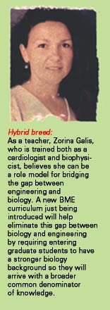
Like Tannenbaum,
cardiologist Zorina Galis segued early training in physical science to
applications in medicine. She says part of her transformation to vascular
biologist was her realization that years spent learning quantum mechanics
and electrodynamics equations could lead to creative solutions to biological
problems.
Galis studies
the mechanisms of matrix metalloproteinases (MMPs)—enzymes that help
cells “remodel” tissue by reshaping their surrounding “scaffolding,”
or infrastructural matrix. As one of the world’s experts on MMPs,
Galis has found that their effects can be either positive or negative.
In cancer, tumors use MMPs to drill holes through the matrix and invade
surrounding tissue. This knowledge led Galis to study atherosclerosis,
in which plaque grows in blood vessels, causing inflammatory cells to
migrate in and proliferate. She discovered that as plaques build up, cells
use MMPs to reshape vascular walls in an effort to repair them.
Although vessel repair starts with the best of intentions, says Galis, there is no cellular switch to turn it off. When MMPs break down the scaffold to allow arteries to enlarge, they improve blood flow but at the same time weaken the plaques, causing them to rupture and block the vessel. She is studying ways to inhibit the harmful actions of some MMPs without stopping the beneficial effects of others. She consults with several companies studying MMP inhibitors to limit restenosis following angioplasty and to stabilize plaques in patients with atherosclerosis. “We have finally reached the point where we understand what drives plaque toward either vulnerability or stability, and the question now is how to stabilize it. This is a major topic now at every cardiology meeting,” Galis says. (BACK TO TOP)
Grafts
of Gortex
Steve Hanson,
another biomedical engineer concerned with the viability of blood vessels,
is well known for his innovative approaches to improving artificial vessels
and developing drugs to prevent and treat thrombosis.
Hanson’s
current research is directed toward patients who need frequent kidney
dialysis. For these patients, access to blood vessels in the arms or above
the knee is an ongoing problem, often necessitating use of artificial
substitutes commonly made from the Teflon-like material Gortex. The typical
dialysis access graft in a patient’s arm lasts only 18 months and
in a leg for several years. Often the grafts cause blood clots or other
problems requiring surgery.
Hanson has patented a method of preserving the life of these artificial grafts by delivering drugs directly into the artificial vessels and into the often troublesome cut edges of the native connecting vessels. The engineered prosthetic vessels have worked well in primate studies, and Hanson has licensed the technology to Durect Corporation. He continues to work with a large group of colleagues within GTEC (the Georgia Tech/Emory Center for the Engineering of Living Tissues) to develop tissue-engineered substitute blood vessels designed from natural materials that could be used for bypass surgery or dialysis. (BACK TO TOP)
Miniscule
pieces of matter
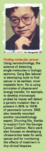
Cancer is
another area rich in opportunities for the blended skills and perspectives
shared by faculty in biomedical engineering. BME faculty member Gang Bao
is using principles of physics and energy transfer to develop microscopic
probes he hopes will detect pancreatic cancer. He believes his probe,
also called a “molecular beacon,” might detect a specific mutation
in a gene that is present in 80% to 100% of pancreatic tumors.
Dr. Bao’s
research is part of the new science of “nanotechnology”—working
with miniscule pieces of matter. The molecular beacon he uses is made
up of a short sequence of single-stranded DNA code synthesized to match
a genetic region where a mutation is known to occur. The beacon uses a
technology called fluorescence resonance energy transfer. When the beacon
finds its target—the mutated gene—the fluorescence is revealed.
Working with
Winship Cancer Institute scientist Lily Yang and Emory surgeon Charles
Staley, Bao hopes his technology can eventually be used to detect a range
of tumor markers, including survivin, a gene expressed in most cancers.
The BME department recently recruited another nanotechnology expert, Shuming Nie, thanks to support from the Georgia Cancer Coalition. Nie is a world leader in detecting single molecules in biologic systems, and his work focuses on developing ultrasensitive tests for detection of cancer at an early stage. (BACK TO TOP)
A
lot of work to do
“Our
department has grown rapidly in a short time, and this is only the beginning,”
says Don Giddens. “We’ve got a new building going up at Tech
and space being renovated at Emory. And BME will more than double its
current faculty, from 12 to 27, by 2005.
“We’ve got a lot of faith and resources vested in us, and all for good reason. Our job now is to to realize our potential through new discoveries that make a difference in people’s lives—and to train the next generation of people who will do likewise, and more so.”
Holly Korschun is director of science communications in the office of Health Sciences Communications.
Friends and forbears:
How the Department Came to Be
1987:
Emory and Georgia Tech established a joint Biomedical Technology Research Center, based on a grassroots relationship among individual researchers at the two schools that had been building for several years.
1990:
The schools' ties were strengthened with creation of the Georgia Research Alliance, a partnership of Georgia's research universities, business community, and state government.
1993:
The Whitaker Foundation awarded Tech a grant to expand biomedical engineering with Emory as partner.
1995:
The two schools established a joint MD/PhD program.
1997:
The two schools formed a shared Department of Biomedical Engineering consisting of two faculty members, including BME Chair Don Giddens.
1998:
The National Science Foundation awarded Tech, with Emory as partner, $12.5 million to establish a tissue engineering center, called GTEC.
2000:
The fledgling BME department attracted a $16 million award from the Whitaker Foundation to add faculty and support infrastructure. In addition, Andreas Kogelnik became BME's first MD/PhD recipient.
2001:
A $25 million commitment from the Wallace H. Coulter Foundation created a new department name and funded faculty chairs, fellowships, and equipment. Perhaps most important, it also created the Coulter Endowment for Translational/Clinical Research, linking biomedical engineers to clinicians on projects leading to direct applications for patients within two to three years, rather than the 10 years or more often required for bench-to-bedside research.
2002:
BME graduated its first PhD student, Andrew Tsourkas. Also, ground was broken for the U.A. Whitaker Building at Tech, and labs and offices are being renovated in Emory's Woodruff Memorial Research Building. By 2005, BME will more than double its current faculty, from 12 to 27.
Copyright © Emory University, 2004. All Rights Reserved