
|
 |
 For better or worse, the anatomy course is a crash course in death: you can't spend sixteen weeks with nineteen cadavers without feeling and pondering death . . . . 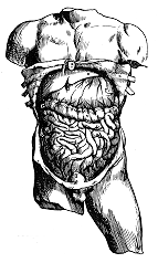 |
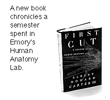
The irony's not lost on Albert Howard Carter III, a professor of comparative literature and humanities at Eckerd College in St. Petersburg, Florida, and author of First Cut: A Season in the Anatomy Lab (308 pp., New York, Picador, 1997. ISBN 0-312-16840-3.) His book is an engrossing and often poetic account of a semester spent at Emory in observing a class of M-1s grapple with the first important rite of passage in medical school, the course in human dissection. "As the anatomy course proceeds," he writes, "I learn many of the students have scant experience with dead bodies; some have never seen one until the first day of lab. For better or worse, the anatomy course is a crash course in death: you can't spend sixteen weeks with nineteen cadavers without feeling and pondering death . . . ." Medicine has been a lifelong source of interest to Carter, who considered it as a career before settling on the humanities and teaching. His fascination with the subject led him to complete training as an emergency medical technician, and earlier this year, Carter released a second book on a medical subject. Rising from the Flames: The Experience of the Severely Burned, written with Jane A. Petro, MD, professor of surgery at New York Medical College, examines the physical, social, cultural, and psychological battles that must be fought by those who have been severely burned. His impetus to write First Cut was a highly personal one - the unresolved issues surrounding the death of his father, also a literature professor, from brain cancer. His father's decision to donate his body to medicine meant no traditional funeral, no last look at the body, no final, private moment. Carter writes about this in his introduction to First Cut: "As my family rallied around [my father] during his long, slow decline, we saw his journey from health to death, the disintegration of his body and mind, the kindness of our family doctor but also the limits of his skills and, of course, the limits of all the other physicians who treated him and could not save his life. My father willed his body for medical research. Upon his death the funeral home sent his body to a medical school some 150 miles away - and that was all we knew. What happened to his body? Whom did it serve? We assumed it was used for the education of future physicians, but never had any definite notion. I wondered for many years what had happened to his body. I'm not a strong believer in the comfort of grave sites or gravestones, but I felt a yearning to know something of 'where he ended up.' Watching these young students begin their journey into the human body, I was searching for my lost father." It was a search that began from the heart, and with the heart. - D.G.
Excerpts from the book |
 or all the violence that permeates American society, and always has, we have become curiously isolated from its logical aftermath. The animals we kill for food are de-boned, re-shaped, and packaged in clean, white, nonbiodegradable Styrofoam. Dead humans are packaged, too, in peaceful poses regardless of their manner of expiration, arms crossed placidly across chests, hair fixed, makeup applied. Gone are the days, for most Americans, of intimate interaction with the dead - of washing, handling, and sitting up with a deceased loved one. We're barraged from birth with violent images in the media and violent realities in our streets and homes, yet the closest most of us actually get to touching death is by pressing 9-1-1 on a cell phone.
  |
 he anatomy books make it sound so simple. To remove the heart, you cut through the pericardia (the two tissue layers around the heart), you make sure the eight major blood vessels are completely severed, and then you lift the heart out of the chest. For the first-year med students in the anatomy lab, however, everything becomes complicated: the first cutting in a crowded area, the hope of a good grade, the fear of "screwing up the dissection," and the thought that this heart was beating, strongly and warmly, in a living human being some weeks ago or even days ago. The med students cut slowly, some taking up to two hours to remove the heart from their cadaver. he anatomy books make it sound so simple. To remove the heart, you cut through the pericardia (the two tissue layers around the heart), you make sure the eight major blood vessels are completely severed, and then you lift the heart out of the chest. For the first-year med students in the anatomy lab, however, everything becomes complicated: the first cutting in a crowded area, the hope of a good grade, the fear of "screwing up the dissection," and the thought that this heart was beating, strongly and warmly, in a living human being some weeks ago or even days ago. The med students cut slowly, some taking up to two hours to remove the heart from their cadaver.
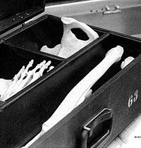
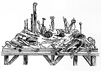
I'm in one of the lab's four rooms to watch these students dissect. In this room there are six tables bearing six embalmed bodies of people who have died in the past year. At each table there are, usually, two students of the Human Anatomy course. At Table 4, however, there is only one student, Karl Jacobs. A quick worker, he is the first to have a heart entirely removed from the body. I watch him cut the last blood vessel and pull the heart up and out. For a moment he stares at it in the palm of his hand, then suddenly raises it over his head in an exaggerated gesture of triumph. Lights flash off his glasses; his lab-coated arm in a dramatic white column. He roars out "Ha-haaaa!" - the very image of a deranged scientist in a Hollywood thriller. Karl makes a huge grimace, as if he were a monster ready to bite right into this grayish but recognizable human heart. Everyone else in the lab stares at this, the first heart in hand and the first truly theatrical gesture of the course. Up to today, students have been sober, timorous, in awe of this place and its strange proceedings. The five other dissecting teams envy Karl's progress and, perhaps, his unabashed progress. "Just like the Aztec priests!" a woman calls out appreciatively from across the room. "Those Aztec guys must have been good - you know, strong," another student says. "To bust through the ribs and sternum, all these tissues." He gestures at the wreckage he is simultaneously creating and ordering in his own cadaver. "Yeah - and fast," another adds. "The victims were alive, remember. The idea was to get the heart out while it was still beating." I shiver briefly, recalling the Diego Rivera mosaics in Mexico City that re-created such primal rites. Although I'm across the aisle at another table, my eyes keep looking at this heart, Karl, and the new cavity in the chest of the spindly dead man lying face up. Now Karl examines the heart, turning it over in his hands. He trims up around it, pats it with paper towels, and starts to identify the coronary arteries. Soon he sets it down and turns to the chest cavity. But he has seen me staring. "Come take a look," he invites. I walk across the aisle to look at this complex thing, meaty, but punched with holes. I lean forward in my starched lab coat, my hands clasped behind me. About the size and shape of a fist, the heart lies on a paper towel. The color of cardiac muscle is somewhere between red and brown, but there are also bands of yellow fat and eight scattered holes, through which I glimpse the four chambers that pump our blood. Under the diagonal bands of fat lie portions of the coronary arteries; I can see them circling down from the top of the heart, vaguely like the "crown" that the word coronary suggests. So, I think, these are the faithful fellows that you don't want to clog with fat. "Go ahead, pick it up: it even fits real well," Karl says. I place my gloved hand over the heart and grasp it gingerly. When it doesn't collapse, I gently tighten my fingers around it. This odd thing pumps in my chest right now, I think. I turn my hand over, and the heart settles into my palm perfectly, just as Karl said. "Hey, you're right," I say, and he smiles, a beginner in anatomy, but already a teacher to me. |
 |
 am surprised when [Professor of Anatomy and Cell Biology] Art English takes the podium and looks apologetic. am surprised when [Professor of Anatomy and Cell Biology] Art English takes the podium and looks apologetic.
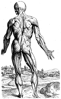
"I'm sorry," he intones, "to have to make this announcement about this morning's lecture, particularly since we take pride in the organization of this course, as complex as it is, but I got a phone call this morning - that the lecturer couldn't make it. Students look at each other. I feel genuine disappointment, since the announced topic, "The Functional Anatomy of the Heart," should be fascinating. "The message was," Dr. English persists, "that Dr. Silverman's athlete's foot was acting up terribly, but that he had found a substitute." Now wait a minute - lots of heads are turning around, as if to ask: Is English on the level? "And when I interviewed this substitute, it appeared to me that he would be up to the task. Actually he knows a lot about it: please welcome Sir William Harvey!" Baroque music wells up from the projection booth and "Sir William" walks majestically down the middle aisle. He is dressed in a seventeenth-century costume, with a large, floppy white collar and a multicolored doublet. He carries a huge tome in his hand, presumably his revolutionary book on the heart, De motu cordis es sanguinis in animalibus (On the Motion of the Heart and Blood in Animals). Our lecturer [Professor of Medicine Mark E. Silverman] assumes the podium and formally begins, "My name is William Harvey and I am four hundred and eleven years old. You'd think Emory wouldn't assign someone of my stature chores in freshman Anatomy, but perhaps it's because I haven't published anything since the seventeenth century." [Laughter] "I'm here to tell you about the most important discovery of all time, my discovery of the circulation of blood in the body. First slide, please." The first slide shows a portrait of Sir William, and the accuracy of Silverman's costume is suddenly clear. Silverman strikes the same pose, and the students applaud. He continues, telling us about Harvey's life, illustrating with slides. One is a map of Europe with a red star for Padua. "I went to Padua - a foreign medical school - not because I couldn't get into one in my own country [appreciative laughter reflects domestic/foreign conflicts in training], but because it was the best in the world, a small southeastern medical school without a football team" [uproarious laughter, as Padua, southeast in Europe according to the map, and Emory University are unexpectedly made parallel]. Silverman goes on to talk about the medical knowledge and methods of the seventeenth century, including the dominance of Galen, the Greek physician of the second century after Christ. "As it was said as late as sixteen forty-nine: 'If dissection differed from Galen, it was because nature had changed!'" It is hard for us in the twentieth century to understand such blind acceptance of authority. Aristotle became a similar model; he gave a number for human teeth in women that was incorrect - when all he had to do was look in Mrs. Aristotle's mouth to get the right answer. Empirical research wasn't his method, nor did anyone challenge him for a long time. Galen worked with animal cadavers and the wounds of Roman gladiators, Silverman continues, and theorized that natural spirits, animal spirits, and the pneuma - or the vital spirit - moved around the body. With the invention of movable type in the West (Gutenberg) and the development of accurate anatomical drawings (Da Vinci and others), the mass media were all set for the revolution of Andreas Vesalius, a native of Flanders, who had dissected animals as a kid [appreciative laughter]. As a grown man, Vesalius stole the bodies of criminals who were hanged outside of Paris, took them home and dissected them, often finding Galen to be wrong. After moving to Padua, he published De humani corporis fabrica (The Fabric of the Human Body)* in 1543, "drawing a savage reaction from those who had bought the wrong book" [huge laughter: the students have paid over $100 for their anatomy texts alone]. . . . "Please raise the screen," he says, and the enormous white screen rises slowly in front of us. It has been so constantly used that I assumed it was permanent. To have it rise majestically seems a rending of the veil. Behind we see the glass screen of a huge television built into the wall. "Let me show you some echocardiography we did this morning." Dr. Silverman punches the controls and a videotape plays on the screen. It's a tape made by ultrasound, showing the heart many times actual size as it pumps. Furthermore, a computer process has colored the Doppler signals of the moving blood red, orange, and yellow, so that there are fluid explosions of dazzling color in the chambers and through the valves, a kind of liquid fireworks in the hypnotic rhythm of a beating heart. It is spellbinding to watch the four chambers compress and expand, the valves between them open and close. "Jesus - he didn't have that last year," one instructor whispers. All one-hundred-plus of the audience stare, mouths agape. "Are you amazed?" Silverman asks. "Yes!" we say with one voice. "Well, that was today, a real patient. She had some blood between the layers of her pericardium, and we had to get it out. Knowing the anatomical structures of the heart is not just a curiosity," he concludes, "but something we use to treat people . . . ." |
 |
t turns out that beside the sink is a good place for me to stand, since a parade of hearts comes to be washed.
"How's your heart?" I ask the students, acutely aware of the ambiguity of "your." They hold up the cadaveric hearts immediately, patting gently with paper towels, eager to show off features they now understand. "Well, ours is pretty small, which made dissection difficult, but here's . . . ." And: "This guy did not follow good health habits, I would say. He cost us a lot of time in getting the fat off of his heart." And: "Look at this: here's his heart attack." Jennifer shows me a coronary artery that wriggles along the top of the heart, then suddenly stops. Below it the color and texture of the heart muscle are different: darker, slacker. Something blocked that artery, perhaps an embolus or a dissecting atheroma. "See, the tissue below all died without oxygen. Killed him." I feel a wave of emotion: we have just seen this man's final fatal secret. |
  |
he living body has weight, volume, multidimensionality. The only ways into its spaces are through ordinary orifices relating to air, food, and sex, or through harmful cuts: wounds. For cadavers, by contrast, cuts are not wounds, but inquisitive gestures, pointed questions (so to speak), or even compliments. There are shy cuts and bold cuts, brief cuts and extended cuts, cuts light as a caress or decisive as a duelist's. Despite these differences, they are all epistemological, all seeking knowledge within the natural order of bodies. This nature, or phusis in the Greek term, becomes the business of the physician: to know the body's parts, their functions, their relations in sickness and in health.
But while we may speak of the anatomy of a flower, a movie actress, or a fetal pig, the true anatomy is not the structure of those actual objects, that material per se, but the discipline, the activity, the intellectual approach that leads to a comprehensive and synthetic wisdom. And much of this emerges from (or at least starts with) the -tomy, the cutting. This cutting shows (and, in a sense creates, by providing consciousness and words) the physical structures. We are not talking about random cutting, or the anatomy course would be over in a few hours of slashing. Nor is it mutilation, through which the symbolic worth of the body is lost; the discipline of anatomy, rightly pursued, provides order, validation, and affirmation, even as the bodies slowly come apart, disintegrating toward chaos. The students I watch seem profoundly touched by the intricacy and majesty of the human body as they painstakingly work their way through it . . . . A cantaloupe vine - like the students, the cadavers, and the lab instructors - has also been my teacher through these weeks of late summer. When my wife and I were moving into our apartment for the year, we parked the rented truck as close as possible to our building but still had to walk around (or through - depending on our patience) a large bed of marigolds. In the midst of them, there was a weedy vine. By what accident had a seed taken root here? A passing bird? The next day, I looked at it more closely and found three cantaloupes attached, two fist-sized, one grocery store-sized. Over the next two weeks, I kept an eye on the volunteer vine and its produce, figuring someone would pick the melon and take it inside, but no one did. One day, I found the melon detached, and an insect inspecting the place where the vine previously attached, presumably the best point of entry. The insect sought to be, in its own fashion, a cutter. With the insect's help, the sun, and various bacteria, the melon would rot, providing nourishment for its many seeds, some of which might bear fruit in their own season. Neither the insect nor the sun would have this fruit, however; I brushed the insect away and took the melon inside to chill in the refrigerator. My wife and I ate it for breakfast; it tasted good. How did the fruit know when to fall off the vine? How did it "cut" itself loose? What complex timings are there for any departures from life? For the turn of the seasons? To what purpose? These are, of course, ancient questions. John quotes Jesus saying, "Truly, truly, I say to you, unless a grain of wheat falls to the earth and dies, it remains alone; but if it dies, it bears much fruit" (John 12:24, R.S.V.). André Gide was captivated by this paradox and entitled his autobiographical memoir after it, Se le grain ne meurt, usually translated If it die. From death comes new life. I begin to think of the cadavers in the anatomy lab as fruits that separated from life, in their own season, each by their own internal necessity. I think of the dead in my family, especially those who died young, including my father. I hope their time was, somehow, right. The med students benefit from the maturity of the cadavers, their ripeness. For everything there is a season, we read in Ecclesiastes, a time to be born, a time to die. Somehow a cantaloupe took root among the marigolds. Somehow each of us appears on earth, children of our parents' egg and seed. And all that is planted eventually dies. Somehow nineteen cadavers, ripe and harvested fruit, are now in this anatomy lab. They are here as fabulous texts, whole libraries of information, ready for the inspection of 113 students, whose cuts are the plowings and plantings that create new knowledge. It is early autumn now in Georgia, the time for fruits to ripen and soon, leaves to fall. Somehow I am lucky enough to be here, to look, to witness. The students cut in the tradition of Vesalius, and I look over their shoulders. We each cut in different ways, and all of us are increasingly able to see more within these bodies and within ourselves, deepening our own humanity while studying the unusual humanity of the dead. |
Dean's Message | In Brief
True Tolerance | Questions of Gender | First Cut
Philanthropy News | Commencement | Alumni News | Class Notes
Copyright © Emory University. All Rights Reserved. Web version by Jaime Henriquez.